More results...

Gas exchange in animals is the biological process through which oxygen is taken in and carbon dioxide is released to support cellular respiration. Different groups of animals have evolved various structures for this purpose, ranging from simple diffusion through body surfaces in unicellular organisms to specialized organs such as gills, book lungs, tracheal systems, and lungs. As animals became larger and more complex, efficient respiratory systems developed to meet their increasing energy demands and ensure the continuous exchange of gases between the body and the environment.
Organisms that perform aerobic respiration take in oxygen and emit carbon dioxide from the body. The gas exchange of the unicellular organisms takes place through the cell wall and the cell membrane.
Organisms that evolved first have comparatively smaller bodies. Therefore, the surface/volume ratio of their bodies is high. Those animals could exchange respiratory gases through their body surface. With the evolutionary process, the size of the bodies of animals is getting bigger. Thus, the surface/volume ratio decreases.
The complexity of the body is also increasing. Because of this, it has increased the energy requirement also. So, it was not very efficient to exchange gases through the body surface. This is why the special respiratory organs have developed during the evolution of animals. Specialized respiratory organs are first clearly seen in the phylum Annelida, where some species developed external gill-like structures.
The surface where exchange of respiratory gases between the body and the external environment is known as the respiratory surface. There are some characteristics of a respiratory surface.
Some organisms do not possess any specialized respiratory organs. The unicellular organisms exchange air through the cell membrane. And some multicellular organisms, such as coelenterates, flatworms, earthworms, and nematodes, exchange gases through the body surface. Other than that, some specialized respiratory organs can be observed in animals.
Gills are a type of respiratory organ that have evolved to exchange gases in aquatic environments. There are two types of gills
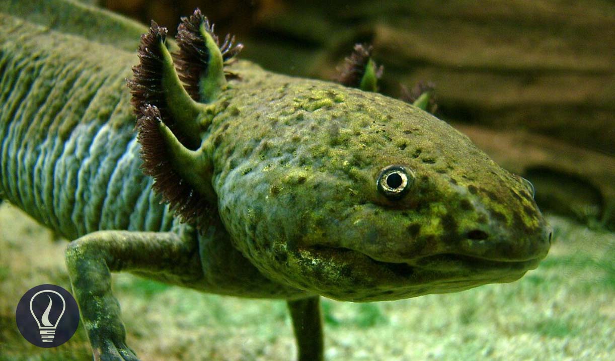
External gills are filamentous structures located outside the body. External gills can be observed as tufts or frilly structures that are attached to the head or body of the organism. Since they are located outside the body, they expose more surface area. It is useful to exchange gases in a low concentration of oxygen. External gills can be found in the larval stage of amphibians, polychaete worms such as Nereis, and Arenicolida.


Internal gills are located inside the bodies of aquatic animals. These are well-protected and efficient structures for underwater respiration. Internal gills can be found in bony fish, cartilaginous fish, and some amphibians.
The internal gill of bony fish is composed of bony gill arches and gill filaments. A large number of gill filaments are connected to the gill arch. These gill filaments act as the respiratory surface. A gill is a well-protected organ, as it is covered with an operculum.
In most fish, water enters through the mouth due to pressure and volume changes in the oral cavity, pharynx, and gill chamber.. When water passes through the gills, it exchanges gases between filaments and water.

Book lungs are a type of respiratory organ found in arachnids such as spiders and scorpions. They are located inside the body and consist of many thin, leaf-like structures called lamellae. These lamellae are stacked together as pages of a book.
Lamella has a blood supply, and they are enclosed in a small chamber. This chamber has a small opening called a spiracle. When air enters the chamber through the spiracle, the oxygen in the air diffuses to the lamella, and carbon dioxide diffuses out.


A tracheal system is the main respiratory organ in insects and some other terrestrial arthropods. The tracheal system of insects is located inside the body. And there is an opening on the body which is known as the spiracle. This spiracle enables air to enter the tracheal system.
The tracheal system is a network of branched tubes. The large tubes are called the trachea, and they are divided into smaller tubes. These tracheae are lined with chitin. The smaller tubes are known as tracheoles. Tracheoles keep dividing into smaller tubes until they reach individual cells. It enables direct exchange of gases between the cells and the air.

Lungs are well-adapted respiratory systems that allow for the exchange of respiratory gases with atmospheric air. Lung systems can be found in terrestrial chordates and aquatic mammals.

The cover image was designed using an image by LoKiLeCh, licensed under CC BY-SA 3.0, via Wikimedia Commons
Figure 01: Contains an image by Florian Witzmann; Elizabeth Brainerd, licensed under CC BY-SA 3.0, via Wikimedia Commons
Figure 02: Contains an image by LoKiLeCh, licensed under CC BY-SA 3.0, via Wikimedia Commons
Figure 03: Contains an image by On the openstax.org page (https://openstax.org/books/biology/pages/39-1-systems-of-gas-exchange#fig-ch39_01_04) where the picture is displayed, it is mentioned the following: "credit "fish":modification of work by Duane Raver, NOAA", licensed under CC BY 4.0, via Wikimedia Commons
Figure 04: Contains an image by John Henry Comstock, licensed under Public domain, via Wikimedia Commons
Figure 05: Contains an image by Original: John Henry Comstock Vector: Pbroks13 (Ryan Wilson), licensed under CC BY 3.0, via Wikimedia Commons
Figure 06: Contains an image by CNX OpenStax, licensed under CC BY 4.0, via Wikimedia Commons